Methaemoglobinaemia is a rare but potentially life-threating condition, with specific clinical and biochemical findings. We present the case report of a woman who became unwell after dental extraction.
This case highlights the importance of adhering to recommended doses of local anaesthetics and recognising the signs of methaemoglobinaemia for timely intervention.
A woman in her sixties with known hypothyroidism, polymyalgia rheumatica and type 2 diabetes was admitted to hospital with hypoxia and syncope. Earlier that day, a dentist had extracted nine of her teeth due to severe periodontitis. Towards the end of the four-hour procedure, the patient was feeling hot and unwell, dizzy and unsteady, and she developed double vision and transient loss of consciousness, which was witnessed by the dentist. The ambulance personnel who attended observed notably low peripheral oxygen saturation (SpO2) of 88 %, despite 6 L/min oxygen supplementation through a mask with a reservoir bag.
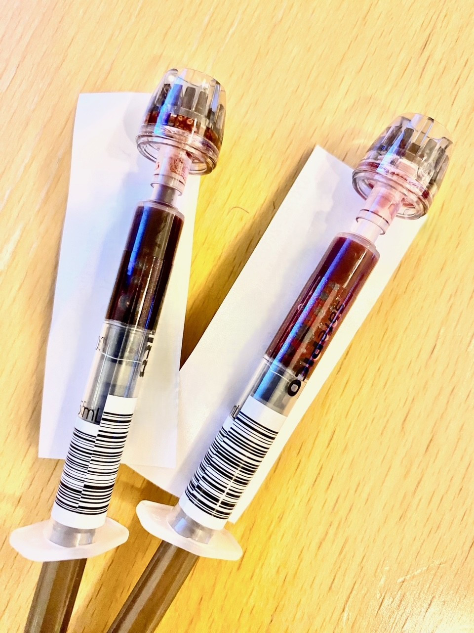
In the Emergency Department, she was awake and lucid, with stable circulation (blood pressure of 127/81 mmHg and regular pulse of 83 bpm) and normal breathing (16 breaths per minute). A review of systems produced normal findings, but she was rather cold peripherally. Low SpO2 of 77 % on room air was noted on finger measurement, while a forehead sensor measured SpO2 of 96 %. The discrepancy was attributed to cold extremities, despite a normal and good saturation curve. The arterial blood gas sample was notably dark in colour (Figure 1), and the analysis results were pH 7.36 (reference range 7.35–7.45); pCO2 4.8 kPa (4.7–6.0); pO2 12.8 kPa (10.0–14.0); HCO3 19.9 mmol/L (22.0–26.0); base excess (BE) −5 (−3 to +3) and lactate 2.5 mmol/L (0.5–2.5). In the Emergency Department, she experienced syncope again with a suspected cardiac cause, but her electrocardiogram (ECG) and blood pressure were normal. However, a more detailed review of the blood gas analysis (Radiometer ABL800 Flex) revealed an elevated methaemoglobin fraction of 25 % (< 1 %), and the patient was diagnosed with methaemoglobinaemia. Since an external cause of the methaemoglobinaemia related to local anaesthesia was suspected, the Norwegian Poisons Information Centre was contacted, and then treatment was initiated with 1 mg/kg intravenous methylene blue (methylthionine) administered over five minutes. The blood sample taken for follow-up blood gas analysis 30 minutes later was considerably lighter, and the methaemoglobin fraction had fallen to 6 % (Figure 1).

The patient was admitted to the medical intensive care unit for observation and received oxygen therapy (9 L/min) through a low-flow mask, with blood gas analysis performed every hour for the first four hours, and then every two hours. She made a rapid recovery, with blood gases returning to normal. A wider case history revealed no similar episodes or other drug reactions in the patient or her close relatives. She was free of symptoms and could go home the day after admission.
Prilocaine remained the only suspected causative agent. The dentist later reported that a total of seven 1.8 ml cartridges of Citanest Dental Octapressin infiltration anaesthesia were administered during the dental extraction, which is equivalent to 378 mg prilocaine. The maximum recommended total dose is 300 mg (1). Prilocaine is an oxidant known to have the potential to cause methaemoglobinaemia. At therapeutic doses, methaemoglobinaemia is rare, although several cases have been reported at doses above the recommended level (2). Citanest Dental Octapressin is contraindicated in patients with known congenital or idiopathic methaemoglobinaemia (1).
Discussion
Methaemoglobin (MetHb) is formed when the iron atom in haemoglobin is oxidised from a ferrous (Fe2+) to a ferric (Fe3+) state that cannot bind oxygen. At the same time, the normal oxyhaemoglobin dissociation curve shifts to the left, which increases its affinity for oxygen, with the result that less oxygen is released to tissues (3–5). This causes blood to be darker in colour, as an indication of the levels of methaemoglobin. Conventional pulse oximetry does not detect methaemoglobin, and at high levels SpO2 is interpreted as approximately 85 % irrespective of the actual oxygen saturation of haemoglobin (6).
The clinical presentation of methaemoglobinaemia is due to hypoxaemia, and clinical features range from weakness, dizziness, headache, 'chocolate cyanosis' (brownish-blue colour/chocolate-coloured blood) and dyspnoea to arrhythmias, seizures, respiratory and circulatory failure, coma and death (3–5). The severity of the condition depends on the level of methaemoglobinaemia, as well as the accumulation rate, the individual's ability to reduce methaemoglobin and any underlying comorbidities (3).
Methaemoglobinaemia should be suspected in patients with discoloured blood (dark red, blueish, chocolate-coloured), particularly when they also present with dyspnoea, chocolate cyanosis and hypoxaemia that is refractory to oxygen therapy, and their arterial partial pressure of oxygen (PaO2) is normal (3–5). The diagnosis is confirmed by biochemistry and blood gas analysis with CO-oximetry, which measures the four haemoglobin derivatives specifically, rather than indirectly using blood PaO2 (4, 6, 7).
The causes of methaemoglobinaemia can be congenital (enzyme defects or abnormal haemoglobin) or acquired (3, 4). Acquired causes of methaemoglobinaemia are most common and include medicinal products or toxic agents, most frequently local anaesthetics, such as benzocaine, lidocaine and prilocaine. Other substances that can induce methaemoglobinaemia are dapsone, chloroquine and nitrites/nitrates (3–5).
Treatment depends on the severity of methaemoglobinaemia and clinical presentation.
Most cases are mild and do not require treatment other than removal of the causative agent (3, 4). High-flow oxygen therapy increases oxygen delivery to tissues and enhances the natural degradation of methaemoglobin (5). Methylene blue is recommended for patients with very high levels of methaemoglobin (above 30–40 %) and patients with lower levels and significant symptoms, particularly those with known cardiopulmonary disease (3, 4). Methylene blue is converted in red blood cells to leucomethylene blue, which reduces oxidised iron (Fe3+) in the haemoglobin back to the ferrous state (Fe2+) that is able to bind oxygen (4, 5). Methylene blue is administered as an intravenous infusion at a dosage of 1–2 mg/kg over 5–10 minutes. Treatment response should be checked 30 minutes after administration, and then every 3–4 hours until stabilisation (8). Due to methylene blue's short half-life, the dose often needs to be repeated, guided by clinical findings and arterial blood gas analyses.
Methylene blue can cause serotonergic syndrome in patients receiving serotonergic antidepressants. Methylene blue should be avoided in patients with known glucose-6-phosphate dehydrogenase (G6PD) deficiency due to the risk of severe haemolysis. In these patients, methaemoglobinaemia is treated with ascorbic acid and, if necessary, exchange transfusion (3–5).
Most patients only require a limited period of observation, but patients with persistent symptoms despite initiation of treatment and/or with severe comorbidities must be admitted to hospital (5). Genetic testing may be appropriate where a hereditary cause is suspected (3). Before discharge, the patient should be given detailed information and advised against future use of causative medicinal products. Adverse drug reactions should also be notified to the Norwegian Medical Products Agency.
A key differential diagnosis is a toxic reaction to local anaesthesia, which is known as LAST (Local Anaesthetic Systemic Toxicity) syndrome. However, apart from syncope, the patient had none of the central nervous system symptoms/signs (seizures, paraesthesia, confusion, agitation) or cardiovascular findings (bradycardia, hypotension, arrhythmia) that are typical of LAST syndrome (9). LAST syndrome was considered unlikely since elevated methaemoglobin was detected and symptoms improved following treatment.
The patient has consented to publication of the article.
The article has been peer-reviewed.
- 1.
Felleskatalogen. Citanest Dental Octapressin. https://www.felleskatalogen.no/medisin/citanest-dental-octapressin-dentsply-547549 Accessed 12.12.2023.
- 2.
Guay J. Methemoglobinemia related to local anesthetics: a summary of 242 episodes. Anesth Analg 2009; 108: 837–45. [PubMed][CrossRef]
- 3.
SWG of red cell and iron of EHA and EuroBloodNet. Recommendations for diagnosis and treatment of methemoglobinemia. Am J Hematol 2021; 96: 1666–78. [PubMed][CrossRef]
- 4.
Solheim L, Brun A-C, Greibrokk TS et al. Methemoglobinemi–årsaker, diagnostikk og behandling. Tidsskr Nor Lægeforen 2000; 120: 1549–51. [PubMed]
- 5.
Ludlow JT, Wilkerson RG, Nappe TM. Methemoglobinemia. Treasure Island, FL: StatPearls, 2023.
- 6.
Annabi EH, Barker SJ. Severe methemoglobinemia detected by pulse oximetry. Anesth Analg 2009; 108: 898–9. [PubMed][CrossRef]
- 7.
Vinnes EW, Holmaas G. Hva består oksymetriparametrene på blodgassutskriften av? Tidsskr Nor Legeforen 2022; 142. doi: 10.4045/tidsskr.21.0855. [PubMed][CrossRef]
- 8.
Felleskatalogen. Metylenblått (metyltionin). https://www.felleskatalogen.no/medisin/forgiftninger/antidoter/metylenblaatt-metyltionin-561453 Accessed 12.12.2024.
- 9.
El-Boghdadly K, Pawa A, Chin KJ. Local anesthetic systemic toxicity: current perspectives. Local Reg Anesth 2018; 11: 35–44. [PubMed][CrossRef]