Gallstones are common, but rarely cause ileus. This case report illustrates the clinical course of a patient who developed gallstone ileus without any previously identified gallstone symptoms.
A man in his eighties was admitted to the department of oncology for biopsy of a suspected malignant lesion in the gallbladder region. He had previously undergone surgery for bladder stones and renal cysts, and had also received hormonal treatment for stage T4 prostate cancer.
Ten days previously, he had been admitted to the department of surgery for observation due to abdominal pain and vomiting. He was diagnosed with COVID-19, which was considered to be the cause. The patient's condition improved and he was discharged without diagnostic imaging. One week later, he was re-admitted to the department of medicine with the same symptoms. Initially, this was also related to COVID-19, but ultrasound of the liver and gallbladder was performed due to elevated liver function tests with aspartate aminotransferase (AST) of 47 U/L (reference range 15–45), alanine aminotransferase (ALT) of 80 U/L (<70) and alkaline phosphatase (ALP) of 115 U/L (35–105).
Ultrasound showed lesions in the liver that were suspected to be metastases. Supplementary CT revealed a suspected malignant mass at the gallbladder and several smaller lesions in the hepatic parenchyma. A wide cholecystoduodenal fistula was also seen, as well as distended small bowel loops in the upper abdomen. The pelvis was not included in the first CT scan, and the level of a potential obstruction was not visualised. The patient was transferred to the department of oncology for a biopsy.
The hepatic lesions were hard to access for biopsy, and therefore the more extensive suspected malignant lesions in the gallbladder region were biopsied. The radiologist who performed the biopsy suspected gallstone ileus on the basis of the CT findings of small bowel dilation and cholecystoduodenal fistula. Ultrasound was used to follow the bowel to a segment with feculent content abutting a round concretion casting a shadow. The gastrointestinal surgeon on duty was contacted, and the patient was transferred to the department of surgery.
On examination, the patient was in a good general condition, with normal vital signs, but complaining of abdominal pain. His abdomen was distended, soft and smooth, with diffuse tenderness on palpation. A nasogastric tube was inserted, which had a good effect on the pain. A further CT scan revealed ileus with a tendency to rotate in the mesentery and calibre variation centrally in the small bowel. However, the gallbladder concretion was not identified on the first examination. Findings of preoperative screening blood tests, including hepatobiliary function tests, were now normal, apart from CRP 121 mg/L (<5). Laparotomy was found to be indicated, and intravenous piperacillin 4 g / tazobactam 0.5 g four times daily was administered due to the risk of bacterial translocation.
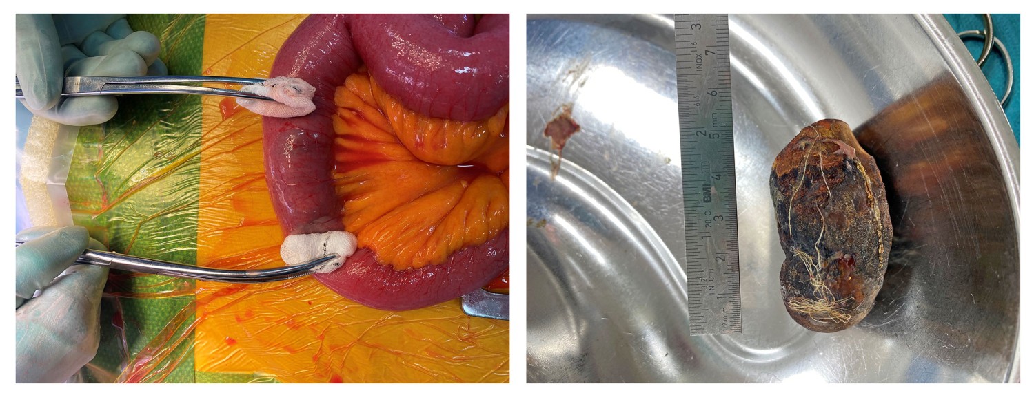
Midline laparotomy revealed an adhesion band from the greater omentum to the small bowel mesentery, which was dissected. An intraluminal structure was identified 200 cm from the ligament of Treitz, and enterotomy was performed and a 5 × 1.6 cm gallstone extracted (Figure 1). The patient made a good recovery and was discharged to his own home three days postoperatively, with further follow-up by the department of oncology. Before discharge, the patient reported having had a gallstone attack over 30 years ago, but at the time it was decided not to perform surgery. He had not had any symptoms since.

The biopsy revealed an adenocarcinoma arising either from the pancreas or bile ducts. The patient was subsequently assessed by the oncologists as only being a candidate for chemotherapy, which he did not want. Therefore, it was decided not to undertake any targeted anti-tumour therapy.
Discussion
Ileus is either caused by mechanical obstruction or bowel paralysis preventing the forward passage of the intestinal contents. These contents accumulate, resulting in bowel dilation. Gallstones are common, with prevalence in Norway estimated to be 22 % in an older study (1), but they cause less than 1 % of cases of bowel obstruction (2, 3).
Our patient illustrates a classic clinical course of gallstone ileus. The concretion has passed through a fistula between the gallbladder and bowel, with fistulas to the duodenum being most common. The concretion advances through the small bowel until it becomes impacted, typically in the distal ileum (3). Prior cholecystitis is thought to be the most common cause of fistula formation (3). Cholithiasis and chronic cholecystitis are also predisposing factors for gallbladder cancer (4).
Gallstone ileus is more common in older people and causes increased morbidity, but mortality rates have fallen to 7 % in the latest studies (2, 3). This may be due to better and faster diagnostic evaluation and perioperative treatment.
Patients with gallstone ileus have usually had symptoms of cholithiasis, but one-third of patients have had no such symptoms (3). Symptoms of gallstone ileus can be acute, intermittent or chronic, and the most common are nausea, vomiting, abdominal pain and abdominal distension (3). An intermittent, subacute course is presumed to be caused by the stone advancing through the gastrointestinal tract until it becomes impacted. Symptoms and findings are related to the severity of obstruction, the size of the stone, and its location. Clinical and biochemical findings can vary from no findings to signs of peritonitis and perforation.
In case of suspected ileus, imaging is usually performed with CT, or possibly ultrasound and/or plain abdominal film. The most characteristic imaging findings are air in the biliary tree (pneumobilia) and detectable gallstone in the bowel lumen. However, radiological diagnosis can be challenging because not all gallstones are radiopaque (3, 5). Most contain cholesterol, and only 10 % contain calcium (6).
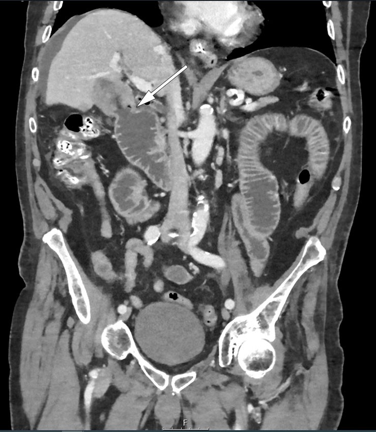
Our patient had a non-radiopaque concretion that was barely visualised on CT. Ultrasound revealed a clear concretion that cast a shadow, and based on the ultrasound findings the outline of this could just be made out on plain abdominal film. A repeat CT scan also revealed air in the biliary tree and cholecystoduodenal fistula (Figure 2). A large concretion in the gallbladder could be discerned on CT images from four years previously, but no fistula or tumour.

Surgery is the main treatment for gallstone ileus, usually with laparotomy, although laparoscopic procedures have also been reported (3, 5). Enterotomy with stone extraction is the preferred treatment (2, 3). Cholecystectomy and closure of the fistula in a one-stage procedure is controversial, but can be considered in low-risk patients (5) or performed later (3). In some cases, the gallstone will pass through the entire small bowel to the colon, so avoiding the need for surgical treatment.
The patient has consented to the publication of the article.
The article has been peer-reviewed.
- 1.
Glambek I, Kvaale G, Arnesjö B et al. Prevalence of gallstones in a Norwegian population. Scand J Gastroenterol 1987; 22: 1089–94. [PubMed][CrossRef]
- 2.
Halabi WJ, Kang CY, Ketana N et al. Surgery for gallstone ileus: a nationwide comparison of trends and outcomes. Ann Surg 2014; 259: 329–35. [PubMed][CrossRef]
- 3.
Nuño-Guzmán CM, Marín-Contreras ME, Figueroa-Sánchez M et al. Gallstone ileus, clinical presentation, diagnostic and treatment approach. World J Gastrointest Surg 2016; 8: 65–76. [PubMed][CrossRef]
- 4.
Wernberg JA, Lucarelli DD. Gallbladder cancer. Surg Clin North Am 2014; 94: 343–60. [PubMed][CrossRef]
- 5.
Ayantunde AA, Agrawal A. Gallstone ileus: diagnosis and management. World J Surg 2007; 31: 1292–7. [PubMed][CrossRef]
- 6.
Schafmayer C, Hartleb J, Tepel J et al. Predictors of gallstone composition in 1025 symptomatic gallstones from Northern Germany. BMC Gastroenterol 2006; 6: 36. [PubMed][CrossRef]