Older patients are often admitted to hospital following a diagnosis of 'acute functional decline'. Assessment typically reveals an acute worsening of a known illness, but occasionally there are unusual causes for the functional decline. This case report relates to a patient with myelitis in the vertebral canal as a cause of falls and acute functional decline.
A woman in her eighties was admitted to hospital with acute functional decline and falls. The patient had been living at home, and was self-reliant and active outdoors. She had a history of peripheral vascular disease and grade 2 chronic obstructive pulmonary disease (COPD), but functioned well physically and cognitively. In the week before admission, the patient experienced a decline in her general condition and an increasing tendency to fall. She described a feeling of weakness in both legs, knee pain and unsteadiness. On the day of admission, the patient fainted and remained lying on the floor, unable to get up. She was confused, and experienced speech latency and dysphasia.
During clinical examination in the emergency department, the patient was afebrile with a temperature of 36.6 °C, blood pressure of 109/84 mmHg and a regular pulse of 76 beats/min. CRP was 7 mg/L (reference range < 4 mg/L) and leukocytes were 9.6 × 10⁹/L (3.5–10.0 × 10⁹/L). Hyponatraemia of 128 mmol/L (137–144 mmol/L) was found, plasma osmolality was 267 mmol/kg (281–295 mmol/kg), urine osmolality was 505 mmol/kg (200–800 mmol/kg) and urine sodium was 55 mmol/L. Renal function was normal with creatinine of 60 µmol/l (45–90 µmol/L). The patient had urinary retention, with 833 ml of urine in the bladder and a positive urine strip test. Hyponatremia was assessed to be due to excessive ADH secretion, known as syndrome of inappropriate antidiuretic hormone secretion (SIADH), and she was treated with fluid restriction. Treatment with pivmecillinam 200 mg × 3 orally for three days was also initiated. The patient was admitted to the observation unit, where she remained for the first 24 hours.
Due to her dysphasia, speech latency and reduced strength in the left leg, a head CT was performed, which showed no signs of acute pathology. Arterial blood gas showed mild hypocapnia 4.1 kPa (4.7–6.0 kPa) and hypoxemia 7.9 kPA (10–14 kPa), and chest CT was therefore performed to assess for pulmonary embolism. Peripheral pulmonary emboli were detected, and the patient was treated with subcutaneous dalteparin 5000 IU × 2.
On day 2, the patient was transferred to the acute geriatric ward for further interdisciplinary evaluation. On day 4, weakness was observed in the left ankle. The following day, a neurological examination revealed pronounced paresis in all joints of the left leg (grade 2/5) and reduced sensitivity to pinprick and temperature sensation in the entire right leg up to the iliac crest and in the distal left leg up to the mid-calf. The patient's knee and ankle reflexes were normal, but an inverted plantar reflex in the left foot and a dorsal response in the right foot were observed, consistent with central paresis in both legs.
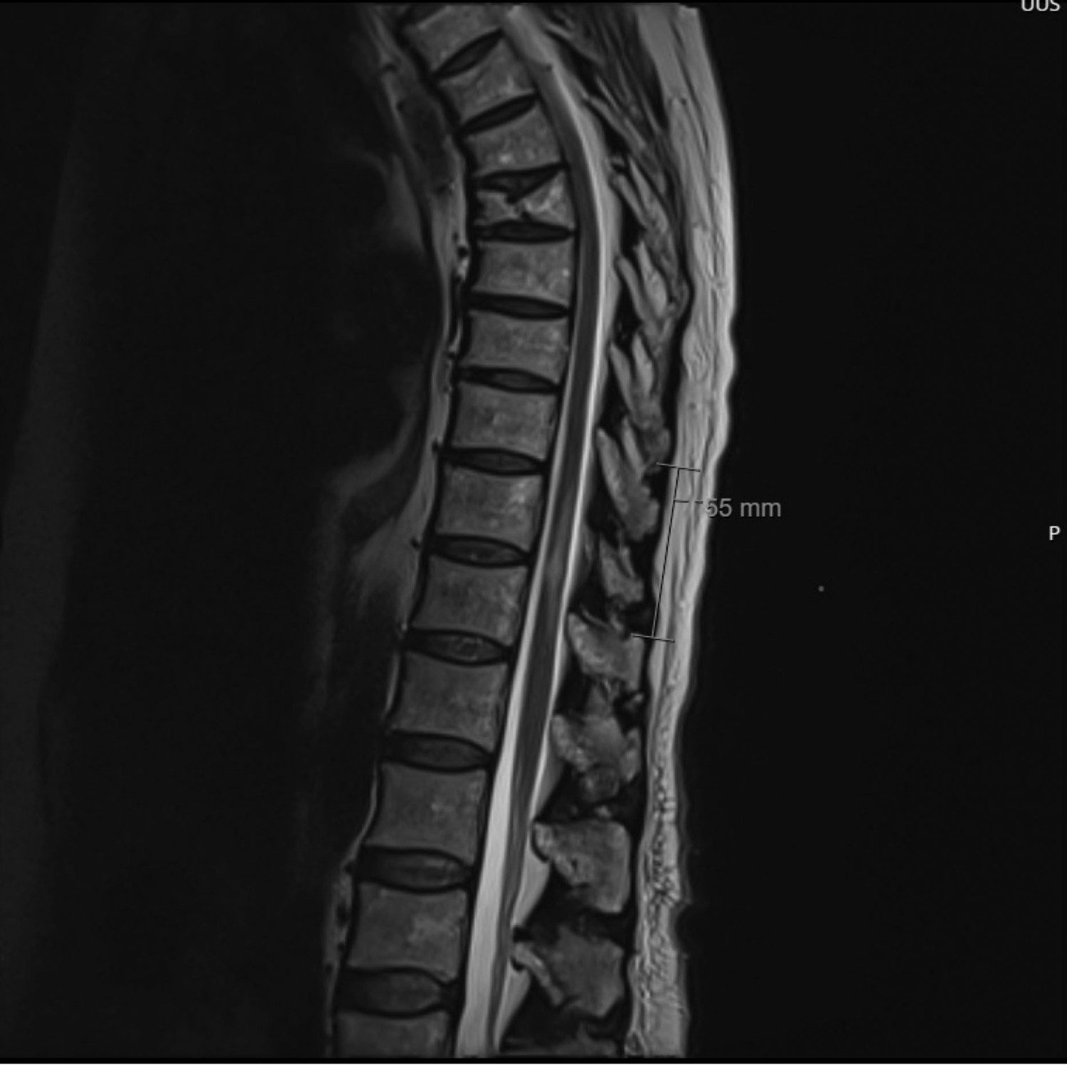
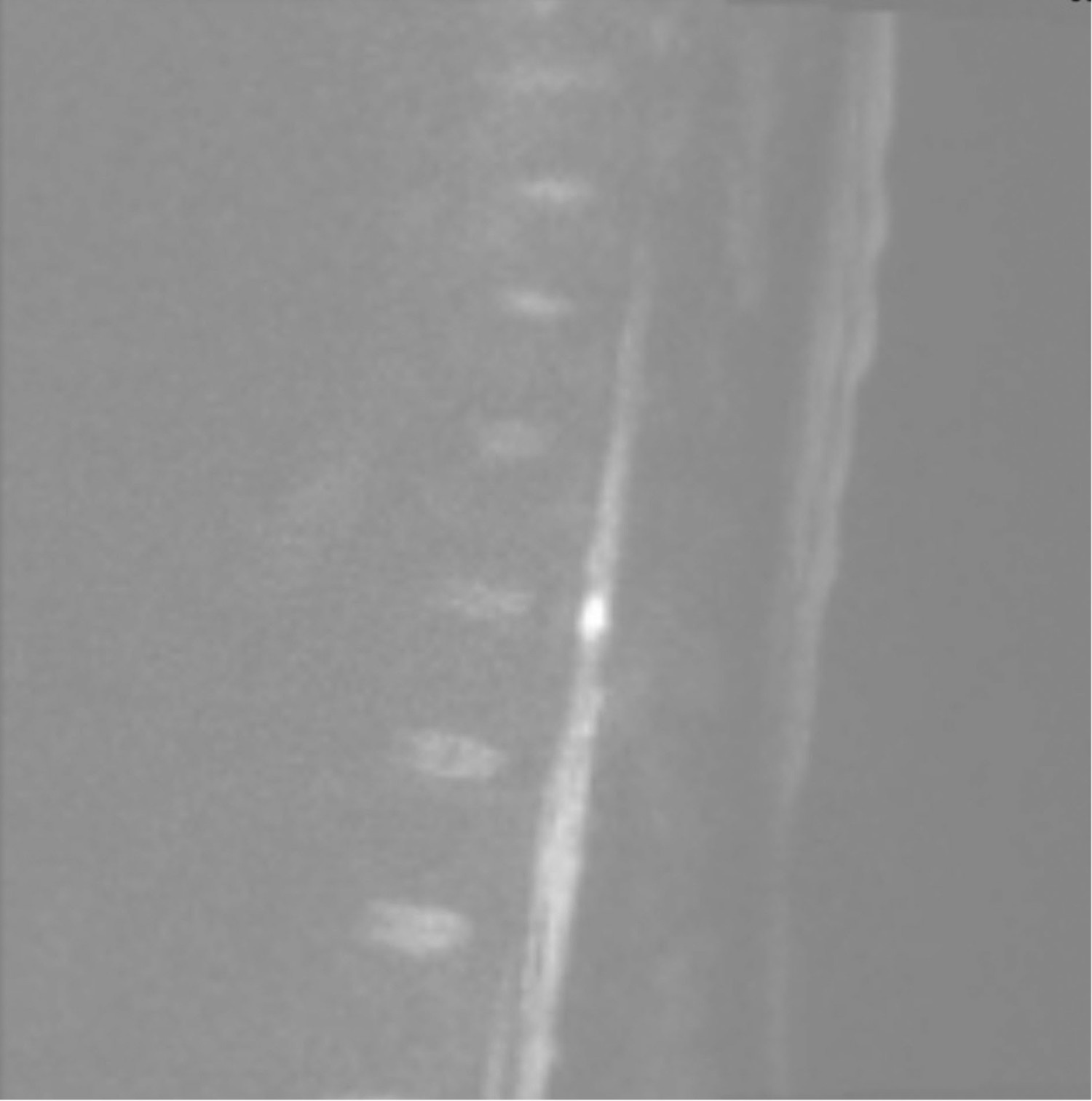
The patient could not be mobilised without assistance and had already been diagnosed with urinary retention in the emergency department. She had now also developed constipation. A head MRI on day 5 showed no signs of acute cerebral infarction. In consultation with the on-call neurologist, an MRI of the entire spinal column was performed on day 6, revealing T2 high-signal changes at the Th11–Th12 levels, measuring 5 cm × 0.6 cm (Figures 1 and 2), consistent with either infarction or myelitis.


The patient was undergoing anticoagulation treatment for pulmonary embolism and could not have a lumbar puncture until three days later. The cerebrospinal fluid was yellowish and cloudy, with 833 × 106/L (< 5 × 10⁶/L) leukocytes. Infectious myelitis was suspected to be the true diagnosis, and treatment was started with intravenous ceftriaxone 2 g × 1 and intravenous acyclovir 1000 mg × 3. Varicella-zoster virus (VZV) DNA was detected in the cerebrospinal fluid on the same day (day 9), and ceftriaxone was discontinued. The patient had no pain. On day 11, she was transferred to the infectious disease department.
At the infectious disease department, the patient's VZV history was reviewed. No rash was detected during her stay, and she had no known history of herpes zoster infection. She was treated with acyclovir for 14 days, without steroids. Repeated lumbar puncture on day 21 showed that VZV was no longer detectable. The patient received daily intensive physiotherapy with mobilisation and rehabilitation.
On day 27, the patient was transferred to the rehabilitation department. On day 30, she experienced acute confusion, incoherent speech and headache, leading to readmission to the neurology department. No new neurological deficits were found upon clinical examination. A head CT showed high attenuation changes in both cerebral hemispheres, and a head MRI revealed multiple small parenchymal haemorrhages. The findings were interpreted as bleeding secondary to anticoagulation. Intravenous acyclovir was restarted at a reduced dose of 750 mg × 2 due to the onset of renal failure during her hospitalisation, with the highest creatinine level at 135 µmol/l. Anticoagulation was stopped due to ongoing bleeding. A new lumbar puncture showed leukocytes at 46 x 106/L, and acyclovir was discontinued after four days when the patient became afebrile, showed no clear new neurological deficits, and an MRI of the entire spinal column showed improvement in the myelitis changes.
On day 35, the patient was transferred back to the rehabilitation department. Upon discharge to a nursing home on day 52, she was able to walk with a forearm walker and could manage stairs. A six-month check-up was arranged with the neurology outpatient clinic.
Discussion
Acute functional decline is a common admission diagnosis for older patients. Functional decline is categorised as acute, subacute or chronic. Acute functional decline refers to an inability to perform daily activities that has taken place within the past two weeks (1). In most cases, acute functional decline is caused by factors such as infection, cardiovascular events, orthostatic hypotension, dehydration and renal failure (2). In some elderly patients, functional decline also manifests with injuries, such as hip fractures associated with falls. The reduction in physical reserves means that the body is unable to compensate when affected by an acute illness. In the field of geriatrics, the term 'frailty' (1, 2) is used to describe the reduced level of functioning caused by factors such as diseases, medications, malnutrition and muscle weakness (3). The degree of frailty can be assessed using, for example, the Clinical Frailty Scale (CFS) (4, 5). The higher the score, the more frail and susceptible the patient is to acute functional decline (3, 4). Our patient had a CFS score of 3 (on a scale of 1–9), which means she has considerable rehabilitation potential despite her advanced age. After a lengthy hospital stay, our patient was self-reliant and could go home. Her acute functional decline was successfully diagnosed and treated through a collaborative effort across multiple specialties and occupational categories.
Myelitis can have various underlying mechanisms, including primary or post-infectious causes related to a range of bacterial, viral and parasitic agents, including enteroviruses or herpes viruses (6). Clinical symptoms typically include sensory deficits and paresis that develop subacutely over several hours to a few days (6). VZV myelitis is an extremely rare condition that can be caused by direct viral infection of the spinal cord (spinal marrow), a post-infectious process or VZV vasculopathy (7). VZV vasculopathy leads to inflammation in the vessel wall, resulting in stenosis and an increased risk of both spinal cord and cerebral infarction (8). The treatment for VZV myelitis is intravenous acyclovir, and the prognosis is relatively good when treated early (9).
The patient has consented to publication of the article and images.
The article has been peer-reviewed.
- 1.
Ranhoff AH. Akutt funksjonssvikt – et vanlig klinisk problem hos eldre pasienter. Indremedisineren 16.4.2014. https://indremedisineren.no/2014/04/akutt-funksjonssvikt-et-vanlig-klinisk-problem-hos-eldre-pasienter/ Accessed 18.6.2024.
- 2.
Wyller TB. Geriatri - en medisinsk lærebok. 1. utg. Oslo: Gyldendal akademisk, 2011.
- 3.
Dejgaard MS, Rostoft S. Systematisk vurdering av skrøpelighet. Tidsskr Nor Legeforen 2021; 141. doi: 10.4045/tidsskr.20.0944. [PubMed][CrossRef]
- 4.
Rockwood K, Song X, MacKnight C et al. A global clinical measure of fitness and frailty in elderly people. CMAJ 2005; 173: 489–95. [PubMed][CrossRef]
- 5.
Legeforeningen. Clinical Frailty Scale. https://www.legeforeningen.no/contentassets/21ef25cf569d44749573de21a8d6b043/cfs_norsk_horisontal_2021.pdf Accessed 11.11.2023.
- 6.
Auwaerter P. Myelitis. https://www.hopkinsguides.com/hopkins/view/Johns_Hopkins_ABX_Guide/540374/all/Myelitis Accessed 17.6.2024.
- 7.
Nagel MA, Gilden D. Neurological complications of varicella zoster virus reactivation. Curr Opin Neurol 2014; 27: 356–60. [PubMed][CrossRef]
- 8.
Kennedy PGE. The Spectrum of Neurological Manifestations of Varicella-Zoster Virus Reactivation. Viruses 2023; 15: 1663. [PubMed][CrossRef]
- 9.
Sebastian AP, Basu A, Mitta N et al. Transverse myelitis caused by varicella-zoster. BMJ Case Rep 2021; 14. doi: 10.1136/bcr-2020-238078. [PubMed][CrossRef]