Infectious ulnar artery aneurysm is a rare condition with no standardised treatment. Our patient was treated with a simple proximal ligature without excision of the aneurysm.
A man in his seventies presented to his general practitioner with declining general condition, dyspnoea and intermittent fever. His medical history included diet-controlled diabetes and asymptomatic atrial fibrillation treated with beta-blockers and a direct oral anticoagulant. Three years previously, he had undergone implantation of a biological aortic valve due to aortic stenosis.
He was treated by his general practitioner for a presumed urinary tract infection with oral pivmecillinam 400 mg three times daily. After five days, this was switched to oral trimethoprim/sulfamethoxazole 160 mg/800 mg twice daily. This also had no effect, and after three days the patient was admitted to the municipal acute care unit. Results of a COVID-19 test and urine test strip were negative, and intravenous cefotaxime 2 g three times daily was initiated. Despite this treatment, the patient had intermittent fever, and after three days he was admitted to the medical department.
Examination in the emergency department found the patient to have pollakisuria and fever. He was in relatively good health, but seemed to lack mental clarity. Findings of auscultation of the heart were normal. Tympanic temperature was 39.4 °C. ECG revealed atrial fibrillation, but otherwise normal findings. Blood tests found haemoglobin 9.5 g/dL (13.4–17.0), CRP 42 mg/L (<5) and normal leukocyte counts. The urine test strip result for red blood cells was 3+. Chest X-ray findings were normal.
The patient was admitted to the department of infectious diseases with a diagnosis of infection of unknown origin, but it was presumed to be a urinary tract infection. He was administered intravenous ampicillin 2 g six times daily and gentamicin dosed according to weight.
Blood cultures taken on arrival showed growth of Enterococcus faecalis, susceptible to ampicillin and vancomycin. Urine cultures showed no bacterial growth. Endocarditis was suspected on the basis of these findings and the fact that the patient had a biological aortic valve. The patient underwent investigation with transthoracic echocardiography after three days, with no findings. Transoesophageal echocardiography two days later revealed a pendulating structure considered to be a vegetation on the biological aortic valve. Small thrombi in the left auricle were also observed.
Following intravenous antibiotic treatment, his CRP decreased from a peak of 45 mg/L to 22 mg/L. The patient was in good clinical condition and afebrile.
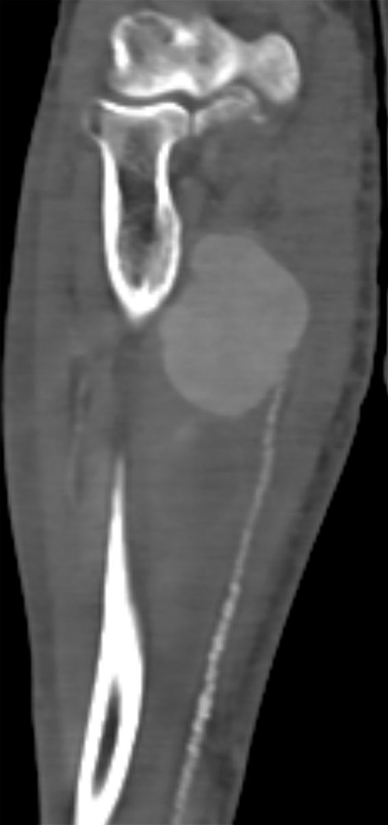
Twelve days after admission, the patient noticed a painful swelling in his right forearm. Ultrasound revealed an aneurysm on the ulnar artery. CT angiography taken the same day confirmed the ultrasound findings and revealed an aneurysm measuring 7.5 × 4 cm (Figure 1). There were numerous atherosclerotic changes distal to the aneurysm. Clinical examination found good circulation to the hand on occlusion of the ulnar artery.

The patient underwent surgery under general anaesthesia for suspected infectious aneurysm secondary to bacterial endocarditis. The brachial artery and bifurcation to the radial artery and ulnar artery were dissected. When we occluded the ulnar artery, the pulse in the aneurysm stopped. We did not dissect the entire aneurysm, but just placed a ligature proximal to the ulnar artery. Following ligation, there was a good pulse in the radial artery, and the colouration and temperature of his fingers were normal.
The patient was administered dalteparin 10,000 + 7,500 IU subcutaneously due to thrombi in the left auricle. There was some bleeding from the skin incision for the first few days postoperatively. This was treated with compression. The bleeding stopped, and the patient reported improvement and decreasing swelling. Ultrasound follow-up on the sixth postoperative day revealed no circulation in the aneurysm.
The total treatment time for endocarditis was planned to be six weeks – the first three weeks with intravenous ampicillin and gentamicin, followed by three weeks of intravenous ampicillin monotherapy. When the patient was discharged to a nursing home for a short-term stay after 32 days, he was being treated with intravenous ampicillin 2 g six times daily.
The patient attended the vascular surgery outpatient clinic 2.5 months after surgery. The pulse in his radial artery was good, but there was no palpable pulse in the ulnar artery. He had no functional symptoms, and strength and sensitivity in the hand were normal. The swelling was considerably reduced.
Discussion
The incidence of symptomatic, peripheral infectious (traditionally called mycotic) aneurysms as a result of infectious endocarditis is 1–5 % (1). The most common locations are intracranial vessels, followed by abdominal vessels and extremities (1). Infectious aneurysms arise as a result of infected emboli in a vascular wall. Surgery is recommended since the weakness in the vascular wall will cause the aneurysm to grow and eventually rupture (2). Several published case reports recommend rapid resection without reconstruction to avoid re-infection, particularly with synthetic material (3).
A review article regarding infected aneurysms in the upper extremities demonstrated that the left brachial artery was most frequently affected, probably due to intravenous drug abuse in right-handed users. Endocarditis was the next most common cause of infected aneurysms in the upper extremities (4). Infectious aneurysms in the forearm have only been reported in single case reports. Most of these are in the radial artery as a result of arterial catheterisation. In the few cases involving the ulnar artery, the main cause was endocarditis, with Staphylococcus aureus being the most common bacteria (4). Among the patients who underwent surgery, all had at least excision of the aneurysm with ligation. A few were treated with bypass and venous graft. None of the patients underwent surgery with only a proximal ligature, which is the procedure we performed.
Our case report indicates that simple ligation of infectious aneurysm in the ulnar artery may be a treatment option in some cases, certainly if it achieves a pulseless aneurysm and there is satisfactory circulation to the hand via the radial artery and collaterals. The patient had a large aneurysm, and full excision would probably have led to faster clinical improvement, but the procedure would have been more extensive and time-consuming and would have increased the risk of complications. Conversely, not excising the aneurysm increases the risk of abscess formation in the aneurysm.
The patient has given consent for the article to be published.
The article has been peer-reviewed.
- 1.
González I, Sarriá C, López J et al. Symptomatic peripheral mycotic aneurysms due to infective endocarditis: a contemporary profile. Medicine (Baltimore) 2014; 93: 42–52. [PubMed][CrossRef]
- 2.
Mieno S, Ozawa H, Tanigawa J et al. A surgical case of excision of infected aneurysm arising from anterior interosseal artery following infectious endocarditis. J Vasc Surg 2011; 53: 1104–6. [PubMed][CrossRef]
- 3.
Prasad R, Handa A. Mycotic Aneurysm of the Ulnar Artery Presenting as a Late Complication of Fulminant Infective Endocarditis. Eur J Vasc Endovasc Surg 2005; 30: 566. [CrossRef]
- 4.
Leon LR, Psalms SB, Labropoulos N et al. Infected upper extremity aneurysms: a review. Eur J Vasc Endovasc Surg 2008; 35: 320–31. [PubMed][CrossRef]