Measurement of high-sensitivity troponin-I in suspected coronary-related chest pain in Emergency Departments
Main findings
The number of admissions to the Emergency Department with suspected coronary-related chest pain was reduced by 31 % after the introduction of an algorithm based on analyses of high-sensitivity troponin I, on admission and after one hour.
For the patients with chest pain, early discharge attributable to the use of the new diagnostic algorithm did not result in significantly increased risk.
Chest pain with suspected acute coronary syndrome is one of the most frequent reasons for patients being referred to a hospital emergency department (ED) (1). The majority are not suffering from acute myocardial infarction, but from a non-cardiac cause of chest pain. The latter are often benign conditions such as musculoskeletal pain or gastrooesophageal reflux (2). Fast, accurate evaluation is crucial to optimal treatment and utilisation of resources.
In earlier European guidelines, a first electrocardiogram (ECG) was recommended within ten minutes of admission, clinical examination and a first troponin test in the ED for patients with chest pain and suspected acute coronary syndrome, followed by a further troponin test and ECG after 8–24 hours (3). Commonly, the patients were hospitalised for diagnostic clarification.
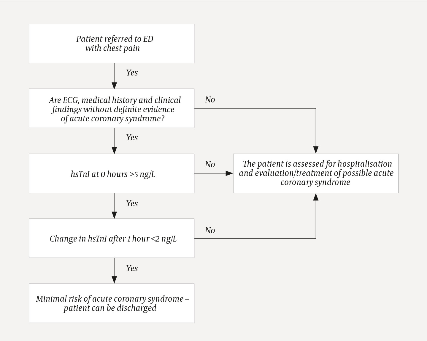
High-sensitivity troponin analyses have paved the way for faster diagnostic rule-in/rule-out of suspected coronary chest pain. The European Society of Cardiology (ESC) recommended in 2015 (4) – with updated guidelines in 2020 (5) – an algorithm for rapid clarification based on a combination of analysing high-sensitivity troponin I immediately after admission and the change after one hour (4, 5). Specific threshold values are recommended in both algorithms for troponin I and troponin T for specified troponin analyses. On 1 March 2017, Levanger Hospital introduced a modified version (the 0 h/1 h algorithm) of the algorithm recommended in 2015 (4) for rapid clarification of chest pain in the ED (Figure 1). It was stressed that in the event of doubt or suspicion of an underlying disease that required hospitalisation, patients should be admitted, even if the requirements for rapid discharge pursuant to the 0 h/1 h algorithm were satisfied. Otherwise, other patients who satisfied the requirements for rapid discharge were discharged directly from the ED. Prior to the introduction of this algorithm, local practice was that the admission troponin I test was performed in the ED, and subsequently at fixed times three times a day at eight-hour intervals.
In order to determine the proportion of patients who were discharged, and any adverse consequences for patients who were referred to the ED with chest pain, and for whom no other cardiac or non-cardiac reason for hospitalisation was found, we compared patient records from a period prior to and a period following the introduction of the 0 h/1 h algorithm.
Material and method
The study was conducted by means of a review of patient records and comparison of samples before and after the introduction of a diagnostic algorithm that used high-sensitivity troponin I for clarification in cases of acute chest pain and suspected coronary disease.
Selection
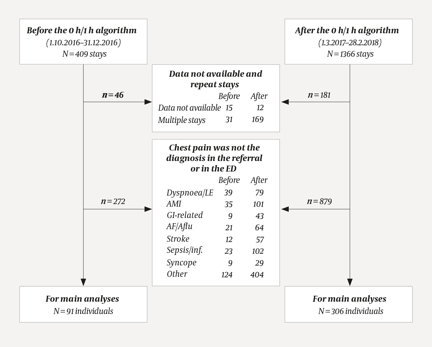
Patients were assessed in the ED as part of routine practice. The samples consisted of patients who were referred to the ED at Levanger Hospital and who had had a high-sensitivity troponin I test. Patient records for the period prior to the introduction of the 0 h/1 h algorithm were retrieved for the period 1.10.2016–31.12.2016. Patient records for the period after the introduction of the 0 h/1 h algorithm were retrieved for the period 1.3.2017–28.02.2018. Data from the patient-administrative system were linked to all high-sensitivity troponin I tests analysed by the central laboratory in both periods. These tests were performed in the ED for a total of 409 patient stays prior to the introduction of the 0 h/1 h algorithm and 1 366 patient stays after the introduction. Repeat patient stays and stays lacking data in the patient records were excluded: 46 from the period before and 181 from the period after.
Patient stays when high-sensitivity troponin-I tests were conducted for a reason other than referral for elucidation of the symptom diagnosis 'chest pain' were also excluded from the material: 187 from the period before and 694 from the period after the introduction.
The resulting groups consisting of 176 patient stays from the period before and 491 patient stays from the period after the introduction of the 0 h/1 h algorithm had all had a high-sensitivity troponin I test due to a diagnosis of chest pain, and formed the basis for analyses of the sample 'referred with chest pain'. After excluding further patient stays where chest pain was not the attending doctor's working diagnosis (85 from the period before and 185 from the period after the introduction), 91 patient stays remained from the period before and 306 from the period after the introduction. These formed the basis for the main analyses. Important reasons for exclusion are shown in Figure 2.
The 0 h/1 h algorithm
In accordance with the 0 h/1 h algorithm (4) (Figure 1) the first high-sensitivity troponin I test was performed immediately after the admission of patients with chest pain which might be due to acute coronary syndrome. After one hour, a further high-sensitivity troponin I test was performed. If the first test was < 5 ng/L, and the test after an hour showed a change of <2 ng/L, the patient was discharged from the ED, provided that the patient history, clinical assessment, ECG, blood tests and chest X-ray (if indicated) did not dictate otherwise. We used a slightly modified version of the European Society of Cardiology's guidelines (4) by always taking two high-sensitivity troponin I tests irrespective of the reading for the first test. The European algorithm recommended direct discharge if the first reading was very low (specified as < 2 ng/L for the assay in question). Our implementation of the 0 h/1 h algorithm entailed clinical evaluation prior to discharge if assay results were normal, as compared to the European guidelines (4).
Analysis of high-sensitivity troponin I
The serum level of high-sensitivity troponin I was analysed with the immunoassay analyser ARCHITECT i2000 SR (Abbott Diagnostics, Maidenhead, England). The lower detection limit was 1.9 (upper measurable level 50 000) ng/L with a 10 % coefficient of variation at a concentration of 5.2 ng/L.
End-points and validation
An experienced cardiologist validated all diagnoses by reviewing patient records for all patient stays in the period before the algorithm was implemented. In the period after the introduction of the 0 h/1 h algorithm, the referral diagnosis and working diagnosis in the ED were first recorded by the attending ED doctor. All other information about the patient stay was validated as described above. Death or acute myocardial infarction (MI) during the follow-up period were validated by another experienced cardiologist through an entry in the electronic patient records. Validation of endpoints took place separately from validation of diagnoses, so that both were blinded for the result of the other cardiologist's validation. The primary endpoint was the proportion of patients with the symptom diagnosis 'chest pain' who were discharged directly from the ED. Secondary endpoints were death or acute MI after 30 days or a year.
Statistical analysis
Normally distributed data are presented as average and standard deviation (SD), non-normally distributed data are presented as median (interquartile range). Proportions are specified as number/total (n/N) and per cent (%). A comparison at group level was performed with Student's t-test for independent samples and the Mann-Whitney U test. Pearson's chi-squared test and Fisher's exact test were used to test proportions. Survival statistics are presented as unadjusted Kaplan-Meier estimates. A Mantel-Cox log-rank test and Cox proportional hazards models (adjusted for age and with/without previous MI) were used to compare the incidence of death and MIs between the samples. Data were censored at 30 days or one year if no event occurred. The statistical significance level was set at p <0.05. All analyses were performed in SPSS.
Ethics
The study was judged by the Regional Committee for Medical Ethics, Central Norway, to be a quality assurance study outside their purview. A legal basis for data processing and exemption from the informed consent requirement were therefore issued by the Nord-Trøndelag Hospital Trust on the basis of the General Data Protection Regulation, the Act relating to Patient Records and the Health Personnel Act.
Results
Samples
Table 1 shows population characteristics for the sample selected on the basis of chest pain as a referral diagnosis and working diagnosis in the ED (primary analysis), and for the sample based on referral diagnosis alone (referred with chest pain).
Table 1
Characteristics of included patients. Characteristics of the sample with chest pain as a referral and working diagnosis in the ED (primary analysis) on the left, and for the sample with chest pain as referral diagnosis (referred with chest pain) on the right. Units are specified.
|
| Chest pain as referral and working diagnosis |
| Chest pain as referral diagnosis | ||
|---|---|---|---|---|---|
| Before the 0 h/1 h algorithm | After the 0 h/1 h algorithm |
| Before the 0 h/1 h algorithm | After the 0 h/1 h algorithm | |
| Total number (n | 91 | 306 |
| 176 | 491 |
| Female, n (%) | 43 (47.3 %) | 147 (48.0 %) |
| 81 (46.0 %) | 214 (44.6 %) |
| Age, mean (SD) | 63.9 (14.1) | 59.7 (16.1) |
| 64.3 (14.7) | 60.8 (16.2) |
| Previous MI, (n (%) | 18 (19.8 %) | 47 (15.4 %) |
| 41 (23.3 %) | 76 (15.5 %) |
| Known atrial fibrillation, n (%) | 3 (3.3 %) | 11 (3.6 %) |
| 12 (6.8 %) | 22 (4.5 %) |
| Troponin I in ng/L, median (interquartile range) | 3 (11) | 3 (6) |
| 4 (27) | 4 (12) |
The average age was significantly lower in the period after the introduction of the 0 h/1 h algorithm (p = 0.03). In the group selected on referral diagnosis alone ('referred with chest pain') the proportion who had previously suffered MI was lower in the period after the introduction of the 0 t/1 t algorithm (p = 0.02).
Hospitalisation frequency before and after
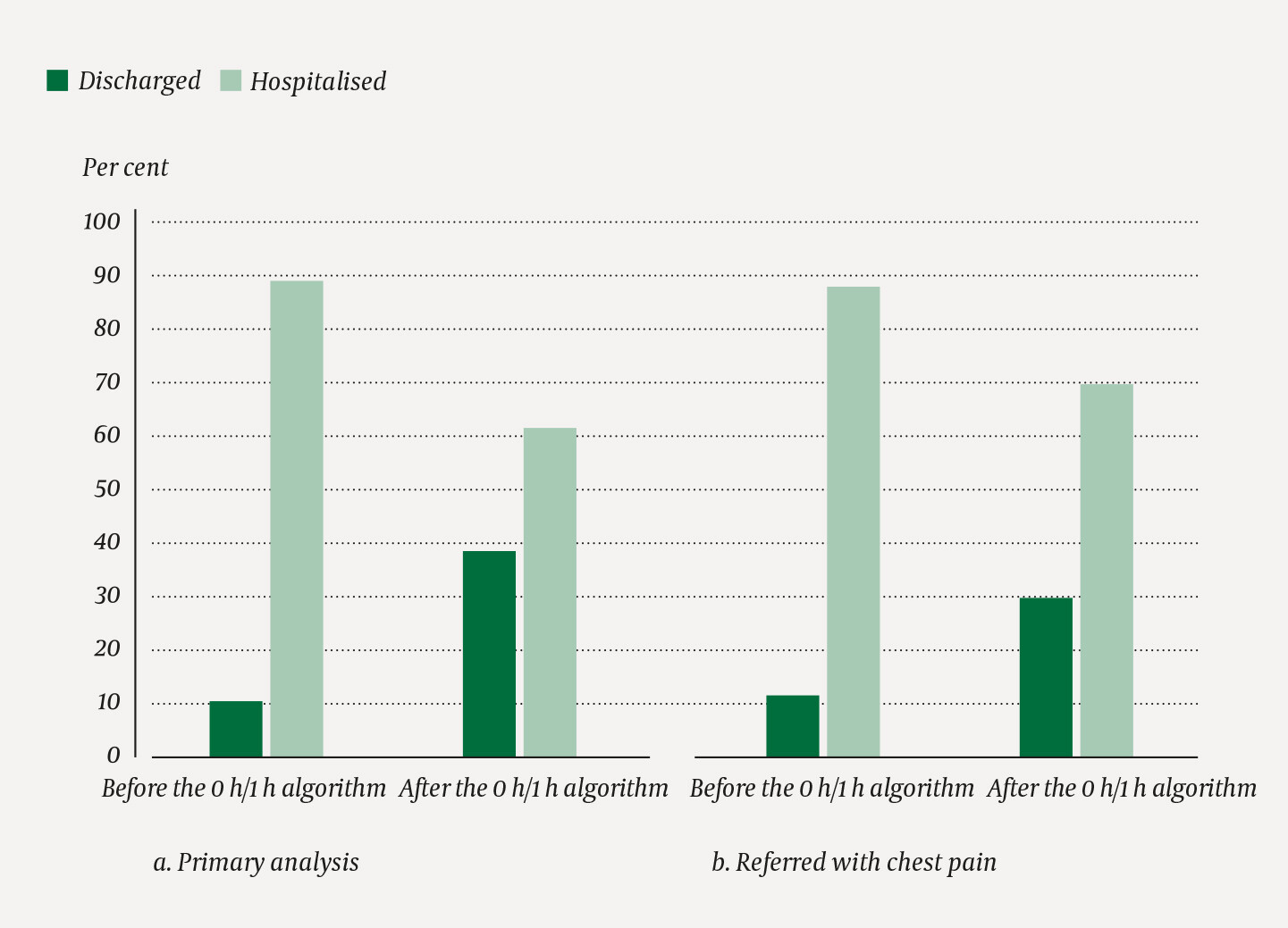
Among patients with chest pain as both referral and working diagnosis (primary analysis) we found that 10/91 (11.0 %) of patients were discharged directly in the period before the introduction, and 118/306 (38.6 %) of patients were discharged directly from the ED in the period after the introduction of the 0 h/1 h algorithm (Figure 3). The relative reduction in hospitalisations was 31 % among those with chest pain as both referral and working diagnosis (primary analysis), and the difference between proportions of hospitalisations/discharges in the periods was statistically significant (p < 0.001). For the sample selected on referral diagnosis alone (referred with chest pain), 21/176 (11.9 %) of patients from the period before the introduction of the algorithm were discharged directly, while the corresponding figure was 148/491 (30.1 %) in the period after the introduction of the 0 h/1 h algorithm (Figure 3). The relative reduction in hospitalisations was 21 % among those with chest pain as referral diagnosis (referred with chest pain), (p < 0.001). Adjustment for age and previous MI, or whether the analyses were performed per patient or per patient stay, did not substantially alter the results (difference between the periods: p < 0.001 (data not shown)). In the adjusted analyses, age was a significant predictor for direct discharge (p < 0.001), but previous MI was not (p = 0.31).
Survival
After 30 days, there were no deaths among those who had chest pain both as referral diagnosis and working diagnosis in the ED (primary analysis) (Figure 4A). At the one-year mark, 3/91 (3.3 %) of the patients from the period before the introduction, and 6/306 (2.0 %) of the patients from the period after the introduction of the 0 h/1 h algorithm had died (primary analysis), p = 0.45) (Figure 4A). Of the patients who were discharged directly from the ED, there were two deaths at the one-year mark in the period after the introduction of the algorithm. These two deaths could not be related to the rapid discharge.
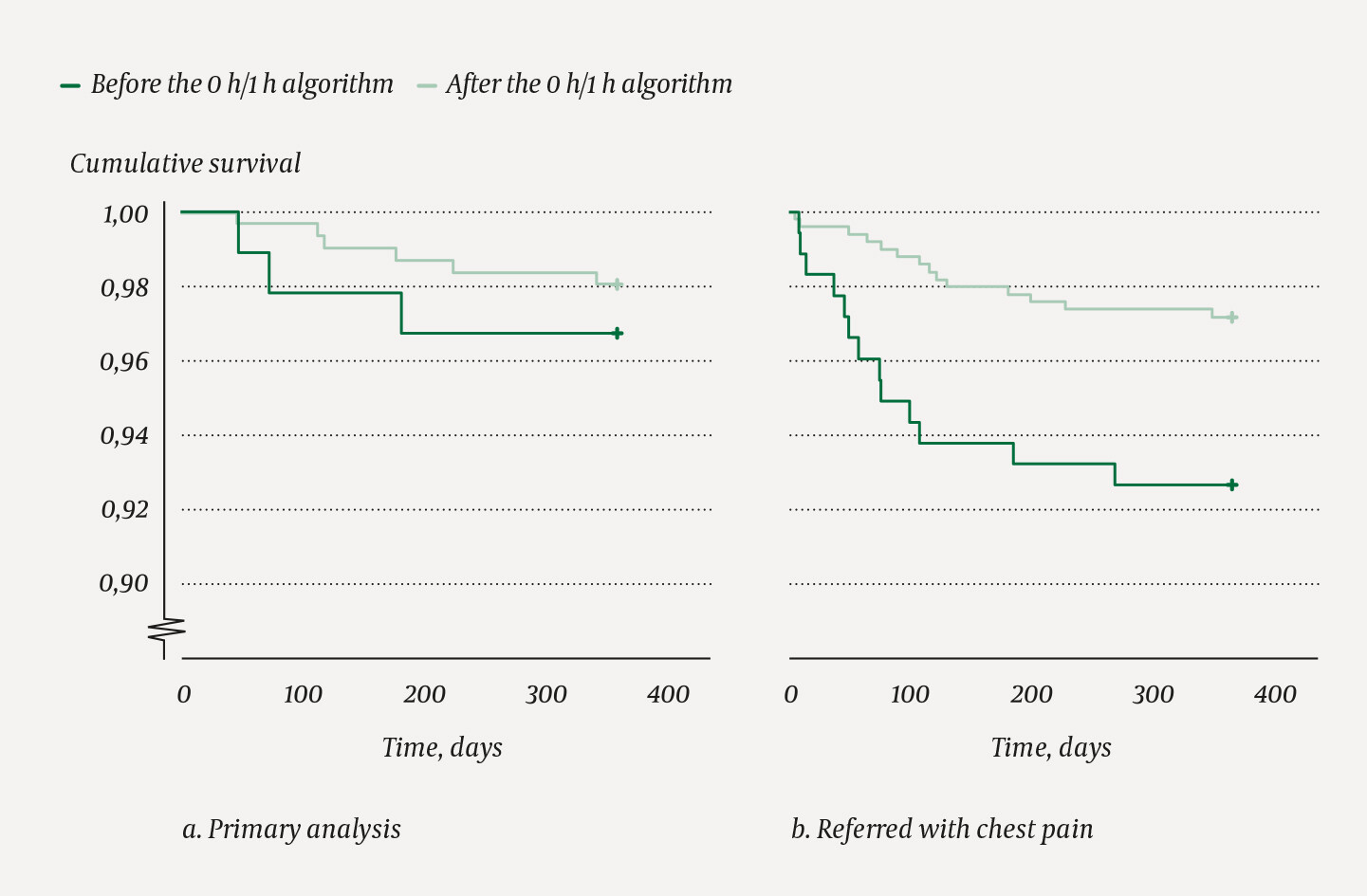
Figure 4B shows mortality when the sample was selected on the basis of the chest pain diagnosis only. At the one-year mark, 13/176 (7.4 %) of patients from the period before the introduction, and 14/491 (2.9 %) of the patients from the period after the introduction of the 0 h/1 h algorithm had died (p = 0.01). None of the deaths could be related to use of the 0 h/1 h algorithm. The difference in mortality between the two periods was not significant after adjustment for age and previous MI.
Acute myocardial infarction
The incidence of acute MI is shown in Figure 5. At the 30-day mark, none of the patients with chest pain as both referral and working diagnosis (the primary analysis), suffered acute MI in the period before, and 1/306 (0.3 %) of patients suffered acute MI in the period after the introduction of the 0 h/1 h algorithm. At the one-year mark, 2/91 (2.2 %) of patients from the period before the introduction, and 2/306 (0.7 %) of patients from the period after the introduction of the 0 h/1 h algorithm had suffered acute MI.
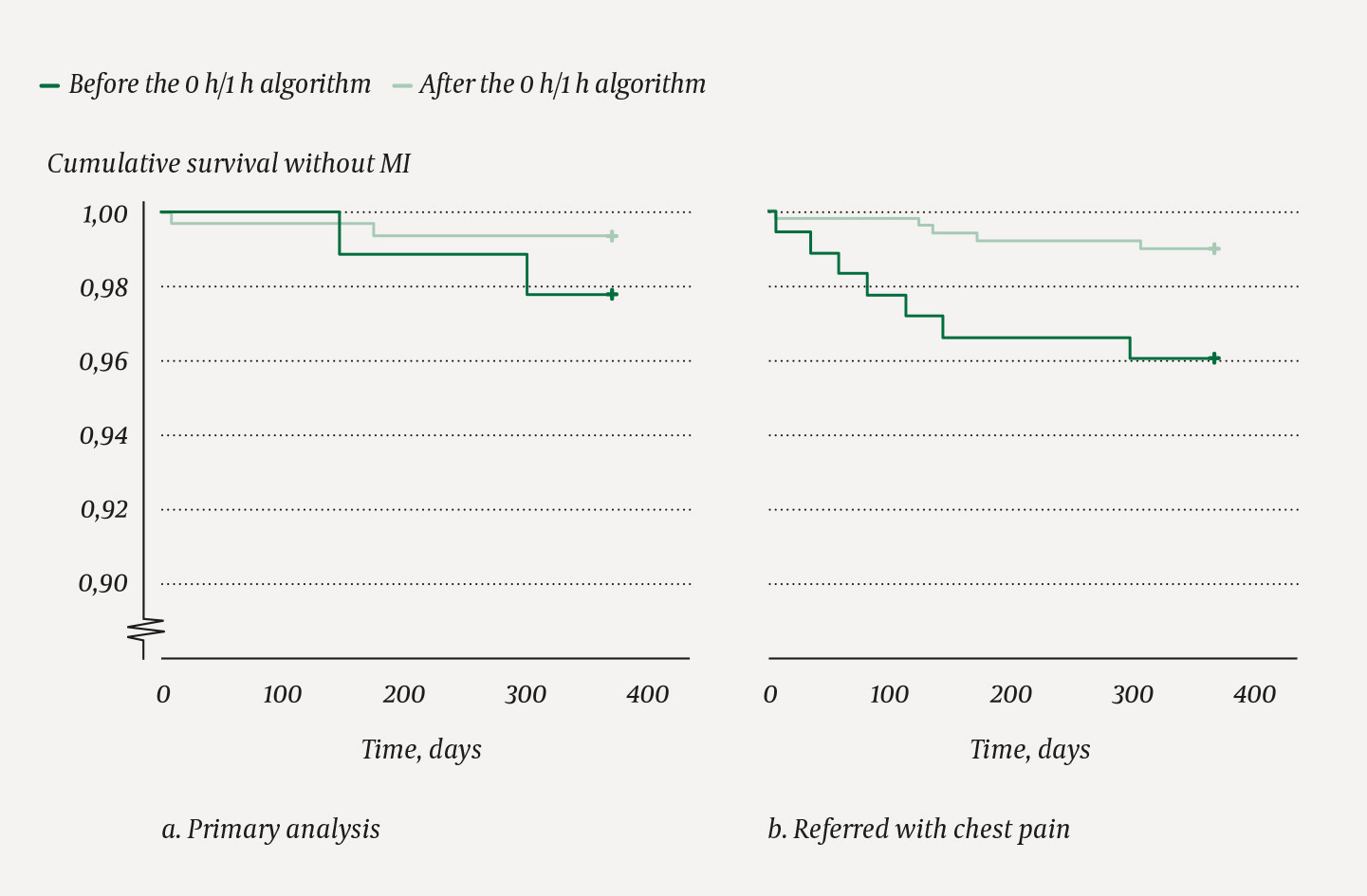
Among those with chest pain only as referral diagnosis (referred with chest pain), 1/176 (0.6 %) of patients in the period before and 1/491 (0.2 %) of patients in the period after the introduction of the 0 h/1 h algorithm suffered acute MI within 30 days. At one year, 7/176 (4.0 %) of patients from the period before the introduction, and 5/491 (1.0 %) of patients from the period after the introduction of the 0 h/1 h algorithm had suffered acute MI (Figure 5B). After adjustment for age and previous MI, there were no statistically significant differences in the number of MIs (all analyses).
Of those who were discharged from the ED, the main analyses showed that 3/10 (30 %) of patients from the period before the introduction and no patients from the period after the introduction of the 0 h/1 h algorithm suffered acute MI within a year (no significant difference after adjustment).
Discussion
Among patients assessed in the ED with chest pain and suspected acute coronary disease, we found a statistically significant relative reduction of 31 % in the number of patients who were hospitalised after the introduction of the 0 h/1 h algorithm. There was no statistically significant difference in the incidence of deaths and acute MI between the samples from the periods before and after the introduction of the 0 h/1 h algorithm after 30 days and one year. None of the patients who were discharged directly from the ED died or suffered acute MI within 30 days.
We conclude that implementation of the 0 h/1 h algorithm for rapid diagnostic clarification of suspected cardiac chest pain appears to represent a safe, effective and resource-saving practice. Several international studies have found the same (6–8). In an international multicentre study with 3267 patients with suspected acute coronary syndrome, the safety and effectiveness of the European Society of Cardiology's algorithm was investigated in a non-selected population (9). The results showed that diagnostic sorting with the aid of the algorithm determined whether the patient had acute MI without ST elevation or not. Prehospital practice and the organisation of emergency departments in other countries are not necessarily comparable to those in Norway. It is therefore important to demonstrate the effectiveness and safety of the algorithm at a Norwegian hospital. The results of our study can probably be generalised to other Norwegian hospitals.
The comparison at group level represents two time periods, and not randomised samples. This has a bearing on the interpretation of the results. The main finding – that a higher proportion are discharged directly from the ED after implementation of the 0 h/1 h algorithm – is judged to be reliable. There were no other changes in organisation or referral practice. We cannot explain the differences by factors other than the introduction of the 0 h/1 h algorithm. The design, seasonal variations in the incidence of illness and admissions and choice of method could affect the comparison at sample level. The period after the introduction of the 0 h/1 h algorithm was longer than the period before the introduction, to ensure an adequate number of events for evaluation of potentially adverse events after the introduction of the new algorithm. The distribution of disease in the samples is fairly even, and we believe that comparison of the samples is relevant, despite the differences in sample size. Patient age was lower in the period after the introduction of the 0 h/1 h algorithm. However, we can conclude with certainty that adverse events such as acute MI and death were very rare after the introduction of the new algorithm.
Chest pain may have other causes than acute coronary syndrome. It is important that patients undergo a thorough clinical examination, and are not triaged purely on the basis of the troponin test. In the event of doubt, the patient's medical history and clinical examination must override the troponin test. In our investigation, the procedure (0 h/1 h algorithm) we introduced in 2017 proved consistent with effective and safe clinical practice. The recommendations of the European Society of Cardiology indicate that different high-sensitivity cardiac troponin assays – including assays for troponin T – can be used (4, 5).
Weaknesses of this study are the retrospective design and different sample sizes. The study has adequate strength for the research question, even though the patient material for examining rare, but serious events such as death and myocardial infarction is relatively limited. Methodological strengths are standardised validation of patient stays, and validation of both the primary endpoint and the other endpoints. We used complete datasets in which all troponin tests from the ED in the periods in question were included, and we used complete follow-up data with respect to the endpoints used. Under-reporting of incidents is theoretically possible, as we did not link the data to national registries. The results are unambiguous, and consistent with what has previously been found in randomised international studies (7), (9–11).
Conclusion
Implementation of high-sensitivity troponin I tests at 0 and 1 hour, coupled with the patient's medical history, clinical examination and ECG, resulted in rapid and reliable determination in patients referred for chest pain. This is consistent with findings from international studies, and can contribute to better patient flow at Norwegian hospitals. A larger proportion were discharged directly from EDs, and we did not experience any increase in the incidence of acute MI or death.
We should like to thank the following for their contributions to the execution of the study: Tina Nordahl and May Shirin Høe, Nord-Trøndelag Hospital Trust assisted with data registration. Bjørn Morten Holm, Central Norway Regional Health Hospital IT, made patient administrative data and laboratory tests available. Marlen Knutli, Research Department, Nord-Trøndelag Hospital Trust, linked patient-administrative data with data generated by the study. The Nord-Trøndelag Hospital Trust financed the study. The article has been peer-reviewed.
- 1.
Niska R, Bhuiya F, Xu J. National Hospital Ambulatory Medical Care Survey: 2007 emergency department summary. Natl Health Stat Report 2010; nr. 26: 1–31. [PubMed]
- 2.
Hollander JE, Robey JL, Chase MR et al. Relationship between a clear-cut alternative noncardiac diagnosis and 30-day outcome in emergency department patients with chest pain. Acad Emerg Med 2007; 14: 210–5. [PubMed][CrossRef]
- 3.
Hamm CW, Bassand JP, Agewall S et al. ESC Guidelines for the management of acute coronary syndromes in patients presenting without persistent ST-segment elevation: The Task Force for the management of acute coronary syndromes (ACS) in patients presenting without persistent ST-segment elevation of the European Society of Cardiology (ESC). Eur Heart J 2011; 32: 2999–3054. [PubMed][CrossRef]
- 4.
Roffi M, Patrono C, Collet JP et al. 2015 ESC Guidelines for the management of acute coronary syndromes in patients presenting without persistent ST-segment elevation: Task Force for the Management of Acute Coronary Syndromes in Patients Presenting without Persistent ST-Segment Elevation of the European Society of Cardiology (ESC). Eur Heart J 2016; 37: 267–315. [PubMed][CrossRef]
- 5.
Collet JP, Thiele H, Barbato E et al. 2020 ESC Guidelines for the management of acute coronary syndromes in patients presenting without persistent ST-segment elevation. Eur Heart J 2021; 42: 1289–367. [PubMed][CrossRef]
- 6.
Lindahl B, Jernberg T, Badertscher P et al. An algorithm for rule-in and rule-out of acute myocardial infarction using a novel troponin I assay. Heart 2017; 103: 125–31. [PubMed][CrossRef]
- 7.
Neumann JT, Sörensen NA, Schwemer T et al. Diagnosis of myocardial infarction using a high-sensitivity troponin i 1-hour algorithm. JAMA Cardiol 2016; 1: 397–404. [PubMed][CrossRef]
- 8.
Shah AS, Anand A, Sandoval Y et al. High-sensitivity cardiac troponin I at presentation in patients with suspected acute coronary syndrome: a cohort study. Lancet 2015; 386: 2481–8. [PubMed][CrossRef]
- 9.
Twerenbold R, Badertscher P, Boeddinghaus J et al. 0/1-hour triage algorithm for myocardial infarction in patients with renal dysfunction. Circulation 2018; 137: 436–51. [PubMed][CrossRef]
- 10.
Reichlin T, Schindler C, Drexler B et al. One-hour rule-out and rule-in of acute myocardial infarction using high-sensitivity cardiac troponin T. Arch Intern Med 2012; 172: 1211–8. [PubMed][CrossRef]
- 11.
Rubini Giménez M, Hoeller R, Reichlin T et al. Rapid rule out of acute myocardial infarction using undetectable levels of high-sensitivity cardiac troponin. Int J Cardiol 2013; 168: 3896–901. [PubMed][CrossRef]